CCHS and Diaphragm Pacing Program
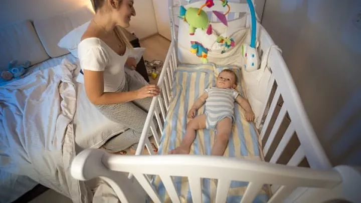
Children born with congenital central hypoventilation syndrome (CCHS) take shallow or slow breaths, especially when sleeping. As a result, oxygen levels drop too low and carbon monoxide levels become dangerously high. Diaphragm pacing is a specialized treatment that regulates your child’s breathing without the need for mechanical ventilation.
The CCHS and Diaphragm Pacing Program at Children’s Hospital Los Angeles is one of the oldest and largest programs in the U.S. We care for a high volume of children with CCHS every year. Our team pioneered a surgical approach for diaphragm pacing that reduces or eliminates the need for mechanical ventilation. This technique is in use worldwide today.
CCHS Care and Diaphragm Pacing: Why Choose Us?
Diaphragm pacing and CCHS services are specialized pulmonology offerings at CHLA. U.S. News & World Report ranks our pediatric pulmonology and sleep medicine services among the country’s best.
Our CCHS and Diaphragm Pacing Program offers:
- Experienced providers: Treating a rare condition like CCHS requires a dedicated care team with specialized expertise. As one of the country’s largest CCHS programs, we see a high volume of children every year. Families from around the world come to us for our CCHS and diaphragm pacing expertise.
- Leaders in innovative approaches: We were the first in the world to place diaphragm pacing electrodes thoracoscopically (using a scope device) through smaller chest incisions. Because we don’t fully open the chest, your child recovers faster and with less pain. Providers from around the world come to us to learn this technique.
- Dedicated CCHS clinic: CCHS affects different body systems. Our CCHS Clinic brings together specialists from various medical fields. Your child sees multiple providers during the same clinic visit, streamlining access to care. Meet our team.
- Care for all ages: There are few CCHS and diaphragm pacing programs for adults. For this reason, our team provides lifelong care for children and adults.
- Family support: We offer supportive services to help your family manage this lifelong condition and the stress that can accompany it. Our social workers, psychologists and other specialists are here for you as you navigate this journey.
Research-focused care: Our team is active in research and clinical trials to advance CCHS care. Your child may benefit from promising new therapy approaches not widely available elsewhere.
What Is CCHS?
An estimated 1,200 people worldwide have CCHS. This rare disorder affects a child’s autonomic nervous system. This network of nerves controls involuntary (automatic) body functions like breathing, digestion and body temperature regulation.
Children with CCHS have a changed (mutated) PHOX2B gene. For most children, this gene mutation occurs for no known reason. In rare instances, a child inherits the changed gene from a parent.
The genetic change affects nerve signals to the brain that control how quickly and deeply your child breathes. Your child’s lungs and heart are healthy, but their brain doesn’t get the signals it needs to regulate breathing. There isn’t a cure for CCHS. However, expert medical care enables most children to grow and thrive into adulthood.
CCHS-related health issues
Children with CCHS may have other health issues, such as:
- Difficulty regulating body temperature, heart rate and blood pressure
- Digestive problems, including Hirschsprung’s disease and bowel obstructions
- Growth hormone deficiency
- Neurological problems, including nervous system tumors (neuroblastoma)
- Vision problems
Expert CCHS Diagnosis
Most children show signs of low oxygen from CCHS—such as blue-tinted skin, fingernails or lips—soon after birth. Experts at our Division of Medical Genetics perform genetic testing (a specialized blood test) to check for the PHOX2B gene change that causes CCHS. You and other members of your family may also take this test. Our medical genetics team offers compassionate genetic testing, counseling and more.
Post-diagnosis tests
After a CCHS diagnosis, your child may get additional tests to assess their overall health:
- Chest CT scan
- Chest X-ray
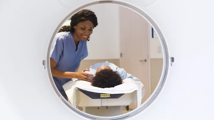
- CT scan, MRI and other imaging tests
- Colonoscopy and other gastrointestinal (GI) tests
- Heart tests, such as an echocardiogram
- Pulmonary function tests
- Sleep studies
What Is Diaphragm Pacing?
Diaphragm pacing is a type of breathing support (ventilation) that regulates your child’s breathing. It can replace or complement mechanical ventilation. Mechanical ventilation requires a ventilator system and tracheostomy (surgically created hole in the windpipe that connects to a ventilator via a tube).
Diaphragm pacing uses surgically implanted electrodes (tiny transmitters) and a small battery-powered diaphragm pacing system. The portable pacemaker sends electrical impulses to electrodes attached to the phrenic nerves, which are essential to breathing. The impulses cause the diaphragm muscles under each lung to expand during inhalation and contract more fully during exhalation.
What takes place during diaphragm pacing surgery?
We evaluate children 2 and older to determine if they’re good candidates for diaphragm pacing. Our general pediatric surgeons and neurosurgeons work together to perform this procedure, which can take up to five hours.
Our team pioneered a thoracoscopic approach, using a scope with a light and video camera to place electrodes. Unlike earlier methods, thoracoscopic surgery takes place through several small chest incisions instead of two large ones. It also doesn’t require spreading the ribs to reach the lungs. This approach causes less pain and scarring, allowing your child to heal faster.
During diaphragm pacing surgery, your child’s surgical team:
- Attaches electrodes to the phrenic nerves on each side of your child’s chest.
- Implants small receiving devices under the skin on your child’s abdomen or upper chest.
- Connects the electrodes and receivers with a lead wire.
- Tests the system to ensure it supports your child’s breathing.
What happens after diaphragm pacing surgery?
After diaphragm pacing surgery:
- Your child recovers in our specialized cardiothoracic intensive care unit (CTICU) for three to five days.
- Your child uses mechanical ventilation support at home for six to eight weeks while their body heals.
- Once they’re recovered, your child returns to CHLA and stays for three days while we initiate diaphragm pacing, adjusting the pacing rate and oxygen levels as needed. You may stay with your child in the hospital.
- To begin, your child uses diaphragm pacing for 60 to 90 minutes a day to give the diaphragm muscles time to get stronger and support deeper breathing.
- The pacing time gradually increases until your child is breathing fully with the pacemaker device (usually within three months).
- You and your child come to our CCHS clinic for follow-up care, as needed.
What are the benefits of diaphragm pacing?
The diaphragm pacing device is small enough to fit into a backpack or fanny pack, making it easy for your child to be active at school, home and with friends.
Diaphragm pacing allows children with CCHS to:
- Be more mobile: Children who need 24-hour breathing support can use diaphragm pacing during the day. Because your child isn’t attached to a mechanical ventilator, they have more freedom to run around and be active. Your child still uses a home mechanical ventilator at night.
- Become ventilator-free: Some children can transition to positive airway pressure (PAP) treatment at night. A PAP face mask delivers oxygenated air. Your child won’t need a tracheostomy or mechanical ventilation system. We carefully screen children to ensure they’re ready for 24/7 diaphragm pacing and PAP.
Multidisciplinary CCHS Care Team
Children with CCHS receive coordinated care from a team of dedicated medical specialists. In addition to pulmonologists and respiratory care practitioners, your child’s care team may include:
- Cardiologists (heart specialists)
- Endocrinologists (hormone specialists)
- Gastroenterologists (digestion specialists)
- General pediatric surgeons
- Neurologists and neurosurgeons
- Sleep medicine specialists
- Ophthalmologists (vision specialists)
- Otolaryngologists (ear, nose and throat specialists)
- Anesthesiologists
Other team members may include:
- Advanced practice providers
- Child Life specialists
- Nurses and nurse care managers
- Physical and occupational therapists
- Psychologists
- Registered dietitian nutritionists (RDNs)
- Social workers
- Speech-language pathologists
Pulmonology and Sleep Medicine Care at Children's Hospital Los Angeles
Our expert team of pulmonology and sleep medicine specialists diagnoses and manages all types of breathing, lung and sleep issues in children. Learn more about our leading-edge Pulmonology and Sleep Medicine services.
Contact us
Pulmonology and sleep medicine experts at CHLA welcome new patients, referrals and second opinions. Please contact us:
- Phone: 323-361-2287
- Online: Make an appointment
- Second opinions: onlinesecondopinion@chla.usc.edu or visit Online Second Opinions