3D Cardiac Printing Enters the Virtual World
For nearly eight years, Jon Detterich, MD, has been printing advanced 3D heart models to help surgeons and cardiologists at Children’s Hospital Los Angeles better plan for complex procedures. But now, Dr. Detterich is leading the hospital’s cardiac 3D printing lab into a new realm: virtual reality.
In December, he and Cardiology Fellow Jennifer Miller, MD, presented their work on virtual 3D heart modeling at the AIMed North America conference in Laguna Niguel, California. The presentation came just two months after the group—which includes computer gaming experts Chaise Allegra and Steven Tu—was one of the winners of the first-ever Digital Health Lab Demo Day, part of the Innovation Studio at Children’s Hospital Los Angeles. The team won $20,000 in seed money to pilot virtual heart modeling at the hospital.
Here, Dr. Detterich talks about how virtual heart models work—and what the future may hold.
What prompted you to investigate virtual reality for heart modeling?
A failed print gave me the idea. A couple of years ago, I printed a heart for one of our surgeons. I had made cuts in it to demonstrate the anatomy where I thought she would want to see it. But when I handed her the model, it wasn’t what she needed.
I brought her into my office, and we made some changes and reprinted it. But I realized that, when you do a 3D print, you’re sort of stuck with it once you print it. Yes, you can reprint it, but that takes time—anywhere from five to 12 hours depending on the size and complexity of the heart.
What is the benefit of a virtual heart model?
In the virtual world, you can manipulate the heart, cut it open and look at it any way you want to. And if you don’t like it, you just click a button to reset it and start over. The results are immediate.
How does it work?
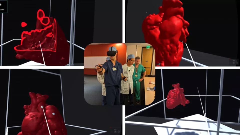
We use the same 3D images from cardiac MRI or cardiac CT that we already use for 3D printing. But instead of printing them, we put them in the virtual world.
The surgeon or cardiologist can then put on virtual reality goggles and see that 3D image. Using hand controllers, they get to grab the heart, twist it, turn it, make rudimentary cuts in it, look inside of it, do anything they want. But then they can also immediately reset everything back to its original state and start over. They’re in control of it; they’re in that virtual world.
Do you see virtual models replacing 3D printing?
Hopefully, we’ll be able to do everything we do with our 3D printed models in the virtual space. Of course, sometimes a physical, printed model will be preferable. Both capabilities are important—using the same images, you can make a print or you can manipulate the model virtually. But the instant access to the virtual models offers a lot of advantages.
What are your next steps?
We’re growing. We were excited to win this grant money from the CHLA Innovation Studio, and now our next step is to build our team and further develop the program.
How do you see the future of virtual heart modeling?
One of my colleagues, Andrew Cheng, MD, is working on computational fluid dynamics using 3D modeling. He’s modeling not only the structures, but also the flow inside the vessels and the heart. Our goal is to get to where we have a virtual model of a beating heart, with blood flowing inside that mimics the cardiovascular system. We’re not there yet. But that’s the ultimate goal.