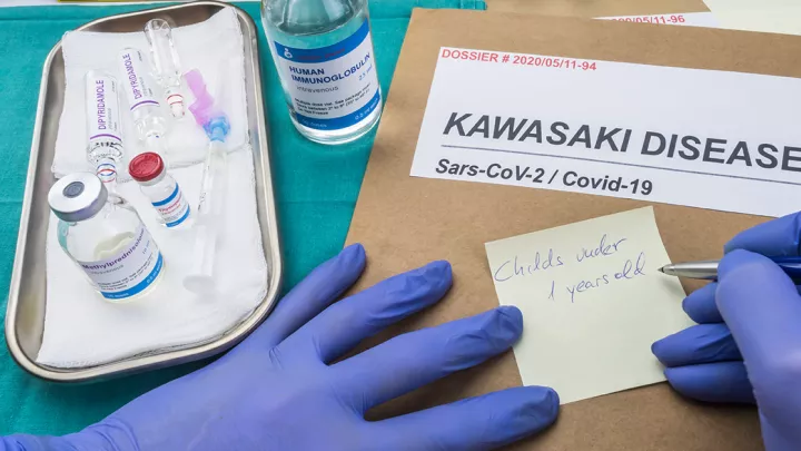
Multisystem Inflammatory Syndrome in Children (MIS-C): What Physicians Need to Know
In March 2020, the World Health Organization (WHO) declared SARS-CoV-2 (Covid19) a global pandemic. Initial data had described the pediatric burden of SARS-CoV-2 as quite low, but by late April 2020, the pandemic epicenters in Europe and the East Coast of the U.S. began to report disease states characterized by varying degrees of multisystem inflammation. Italy, the United Kingdom and New York reported their observations on this emerging phenotype as ranging from “Kawasaki-like” to “hyperinflammatory shock” and our experience at Children’s Hospital Los Angeles has been consistent with these reports. In April and May 2020, we saw a remarkable uptick in patients presenting with features of Kawasaki disease (KD) and incomplete KD, with varying degrees of shock.
|

On March 14, 2020, The Centers for Disease Control and Prevention (CDC) described the condition and named it multisystem inflammatory syndrome in children, or MIS-C, but it has been reported under other names and acronyms (pediatric inflammatory multisystem syndrome, PMIS, PIMS, PIMS-TS, etc.). Though the phenotype is not yet well categorized, it is emerging as a post-infectious complication of SARS-CoV-2 in the pediatric population.
It is crucial to remember that MIS-C remains an emerging phenotype, and the CDC and WHO case definitions are purposefully broad and inclusive for this reason. A new symptom or sign will not make its way into the case definition until astute physicians report it as such! As pediatricians on the front lines, we are faced now with the great challenge of looking for a new kind of needle in the haystack, armed only with a vague and evolving description it. We look to see if a patient fits into the broad CDC template, and then grapple with those clinical features that do not fit; do we dare to report them as “new” features of MIS-C, or do we lean into an alternative plausible diagnosis?
Ultimately, the big question we all have is: What are the consequences of under-diagnosis or over diagnosis of this condition? We must avoid unwarranted alarm and stress, but can we be sure we are not missing permanent coronary artery changes? We struggle with questions such as:
- Fever for how many days, and how high before we check labs?
- Does a skin rash and conjunctivitis count as one or two organ systems?
- What is the trigger to check labs for inflammation or for evidence of SARS-CoV-2 exposure?
- For Patients with incomplete or atypical findings of KD, when do I refer to cardiology to look for coronary artery involvement?
- Is this a continuous spectrum of a disease process, or are there discrete phenotypes? Can a relatively well-looking febrile child progress to shock?
- Can a relatively well child with fever and some signs of inflammation progress to develop coronary artery aneurysms in the absence of other KD features?
Dissecting the case definitions further:
- Fever: The case definition states “³38° for ³ 24 hours, or subjective fever ³ 24 hours.” We have seen reports from 1 to 10 days, and the range from 101° to 105°. It is not necessarily continuous and unrelenting; we have seen patients arrive to medical care afebrile, and then mount a high fever shortly after.
- Gastrointestinal: Abdominal symptoms such as discomfort and diarrhea are very common; rarely it may reach the level of surgical abdomen, and patients with a negative exploratory laparotomy have been reported.
- Conjunctivitis: This is not mentioned above, but is common. It may be short-lived, and can occur early or late in the febrile illness, so it can be part of the history and not a physical sign.
- Dermatologic: Skin rash is variable, and we have seen it range from a minimal macular rash on one hand to extensive polymorphous blanching lesions throughout the body. It can occur at any point in the illness, and so may be historical rather than a diagnostic sign.
- Cardiac: Cardiac involvement has been reported as transient pericardial effusion and/or decrease in myocardial function signaled by hypotension, cardiomegaly or abnormal BNP or troponin. Of particular concern is the development of coronary artery dilation and aneurysm formation, but these can only be evaluated by meticulous echocardiographic examination by highly skilled pediatric sonographers.
- Vascular: This is not singled out in the definition, but vasculitis and vasoplegia-induced hypotension are a frequent feature in many patients. This has been reported variably as a slow decline in blood pressure, or as rapidly progressive hypotension occurring suddenly during the course of the illness.
- Respiratory: The lungs have not, to my knowledge, been reported as a primary inflammatory focus, but pulmonary edema as a consequence of heart failure and pleural effusions secondary to fluid management for hypotension have been commonly reported.
- Renal: This has been rare and associated with severe shock.
- Neurological: This has been infrequent, and reported as severe irritability, confusion, encephalitis or sterile pleocytosis. Stroke has been rare.
- Hematologic: Signs of hypercoagulability with end-organ ischemia have been rarely reported.
- Lab evidence:
- Inflammatory markers are all elevated, including CRP, ESR and ferritin.
- BNP and troponin may be elevated as part of myocardial dysfunction or fluid overload.
- Leukocytosis is common, curiously in combination with anemia, thrombocytopenia and lymphopenia.
- Patients with typical KD may have thrombocytosis. Patients may have elevated d-dimers and elevated fibrinogen.
- Hypoalbuminemia is common; other liver function tests, pancreatic enzymes and renal function tests have tended to be normal.
- Evidence of relation to SARS-C0V-2: The inclusion of evidence of SARS-CoV-2 infection, by PCR, serology, or simply possible exposure, is different from the original definition proposed on May 1, 2020 by the RCPCH, and this deserves some thought and discussion. Many patients are indeed serology-positive (IgG) with no history of exposure, but many clinics do not have access to serology testing. In addition, it is possible that in some patients where the antibody test is negative, it might be in the process of evolving. IgM and rectal swab testing might fill the time gap between PCR-positive and IgG-positive tests, but these are not widely available. The CDC and WHO recommendations, therefore, amount to an amorphous “index of suspicion.” Fortunately, gold-standard testing and expertise is available through Children’s Hospital Los Angeles.
Diagnosis:
Patients come to medical attention via different pathways, and every child should continue to receive the best available treatment as determined by the medical team. With a history, physical examination and laboratory evidence of MIS-C, an echocardiogram is indicated to look for coronary artery involvement, as this may change treatment.
A multidisciplinary team at CHLA cares for these patients. This includes Hospital Medicine, Intensive Care, Emergency Medicine, Laboratory Medicine, Cardiology, Infectious Diseases, Rheumatology, and Clinical Immunology and Allergy. Additional teams may be involved based on the clinical findings.
Treatment:
With all patients, the cornerstone of therapy is astute observation to identify all aspects of their clinical syndrome, with supportive care and customized subspecialty care as needed.
For patients who appear to have KD or incomplete KD, IVIG and moderate dose aspirin is added to their treatment.
When there is an inadequate response to IVIG, other medications may be used, including methylprednisolone, infliximab, anakinra or tocilizumab. The coronary arteries are followed very closely and meticulously with echocardiography, as this guides the anti-thrombotic treatment. For example, medium aneurysms are treated with aspirin and Plavix, but giant aneurysms are treated with aspirin and enoxaparin or warfarin.
For patients who are hypotensive, it is important to assess their cardiac function and support early with vasopressors, as they are often vasoplegic and not responsive to IV fluids.
MIS-C patients frequently have other medical problems that require attention, and there have been reports of concomitant lymphadenitis, UTI, diabetes and pancreatitis, to mention a few examples.
Conclusion:
We here on the West Coast of the U.S. are experiencing the SARS-CoV-2 pandemic slightly after the East Coast and Europe. This is a double-edged sword. On the one hand, it is very helpful to have timely anecdotal data from colleagues across the globe to anticipate and plan the best treatments for our patients. On the other hand, as the data is incomplete and evolving, we are left with uncertainty about applying this information in a rigid manner. There is a feeling of anxiety and uneasy anticipation, caught between wanting to follow clear guidelines, and realizing that the guidelines are far from clear and are evolving. To avoid being trapped in this dilemma, and to avoid cognitive bias, it is crucial to have a high index of suspicion for disease relating to this novel condition when evaluating children with fever and signs of inflammation, and in those who have unusual symptoms or severity compared to the expected course for the presumed diagnosis. Our knowledge of what may be encompassed by this syndrome is still evolving. Utilizing the advanced notice we received from our global colleagues, the iterative process of communicating our own observations and management will be key to clarifying the phenotypes and developing best practices.
References:
- World Health Organization (WHO). Director-General's opening remarks at the media briefing on COVID-19 - 11 March 2020. https://www.who.int/dg/speeches/detail/who-director-general-s-opening-remarks-at-the-media-briefing-on-covid-19---11-march-2020
- Dong Y, Mo X, Hu Y, et al. Epidemiological characteristics of 2143 pediatric patients with 2019 coronavirus disease in China. Pediatrics. 2020; doi: 10.1542/peds.2020-0702
- Lu X, Zhang L, Du H, Zhang J, Li YY, Qu J, et al. SARS-CoV-2 infection in children. N Engl J Med. 2020. https://doi.org/10.1056/NEJMc2005073.
- Ludvigsson JF. Systematic review of COVID-19 in children shows milder cases and a better prognosis than adults. Acta Paediatr. 2020 Jun;109(6):1088-1095. doi: 10.1111/apa.15270.
- Coronavirus Disease 2019 in Children—United States, February 12–April 2, 2020. MMWR Morb Mortal Wkly Rep. 2020;69:422–426
- Verdoni L, Mazza A, Gervasoni A. An outbreak of severe Kawasaki-like disease at the Italian epicentre of the SARS-CoV-2 epidemic: an observational cohort study. Lancet. 2020. doi: 10.1016/S0140-6736(20)31103-X.
- Riphagen S, Gomez X, Gonzalez-Martinez C, et al. Hyperinflammatory shock in children during COVID-19 pandemic. Lancet 2020. doi: 10.1016/S0140-6736(20)31094-1
- Jones VG, Mills M, Suarez D, et al. COVID-19 and Kawasaki Disease: Novel Virus and Novel Case. Hosp Pediatr 2020. doi: 10.1542/hpeds.2020-0123.
- Centers for Disease Control and Prevention. Health Alert Network (HAN) Health Advisory. Multisystem inflammatory syndrome in children (MIS-C) associated with coronavirus disease 2019 (COVID-19). May 14, 2020. CDCHAN-00432. https://emergency.cdc.gov/han/2020/han00432.asp
- O'Sullivan ED, Schofield SJ. Cognitive bias in clinical medicine. J R Coll Physicians Edinb. 2018;48(3):225‐232. doi:10.4997/JRCPE.2018.306
- Viner RM, Whittaker E. Kawasaki-like disease: emerging complication during the COVID-19 pandemic. Lancet. 2020;10.1016/S0140-6736(20)31129-6. doi:10.1016/S0140-6736(20)31129-6
- RCPCH report on inflammatory illness at https://www.rcpch.ac.uk/news-events/news/leading-paediatricians-publish-case-definition-illness-affecting-children-during