Breath In, Branch Out
Inadequate lung development can lead to a variety of pediatric disorders, especially in babies born prematurely. Understanding what drives proper lung development is important to developing new therapies for children with these disorders, but researching this can be complicated because the conclusions derived from studies in animals do not always cross over well to humans.
“Over the last decade, a lot of data emerged from animal studies that may or may not be applicable to humans. Hence, our goal is to focus on what drives proper lung development in the human lung,” says Denise Al Alam, PhD, principal investigator in the Developmental Biology and Regenerative Medicine Program at The Saban Research Institute of Children’s Hospital Los Angeles whose laboratory studies the branching of airways in the developing lung.
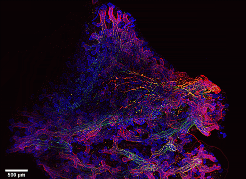
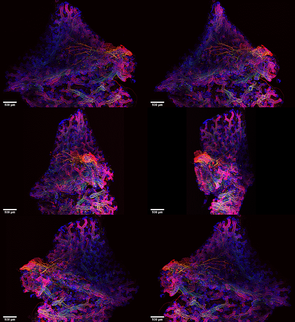
Here we see branching in a 10-week old human fetal lung. Using fluorescent antibodies, researchers imaged epithelial cells along the branches in the lung (blue), smooth muscle cells (red) and nerve cells (green). With a confocal microscope, they were able to generate this 3D image of the lung.

“We see here an intriguing pattern of smooth muscle cells where they form a knot in the middle of the epithelial buds, forming a bow-tie like pattern,” explains Al Alam. “Additionally, previous studies have shown that nerve cells are important for branching in mouse lung, though the pattern and distribution of nerve cells in the developing lung are yet to be described. Here we show the presence of nerve cells extending along the smooth muscle cells to the very tip of the epithelial branches, suggesting an important role for these cells in branching morphogenesis.”
Al Alam and her team are continuing to investigate this topic to determine the specific roles of smooth muscle cells and nerve cells and their interactions in proper lung development.
Images courtesy of Denise Al Alam, PhD, and Esteban Fernandez, PhD, The Saban Research Institute , CHLA.


