Babies and Soldiers: Detecting lung injury
Military personnel can be exposed to dusts and toxicants in the field that may contribute to lung disease. The available technologies to detect early-stage alterations of lung function in patients require a long processing time and lack satisfactory sensitivity and resolution, according to a team of researchers at Children’s Hospital Los Angeles (CHLA). That team has been awarded $2.3 million by the Department of Defense to develop an innovative endoscopic device for performing noninvasive, quantitative analysis of lung epithelial cell metabolism during lung injury.
Their goal is to build a new fiber-optic system that can be used alone or as a modification to an existing upper or lower airway scope instrument, that will detect airway lining cell injury with a greater specificity, sensitivity, and speed than can be done with existing medical technologies. The product of this research is anticipated to significantly improve the respiratory health of military personnel in the field.
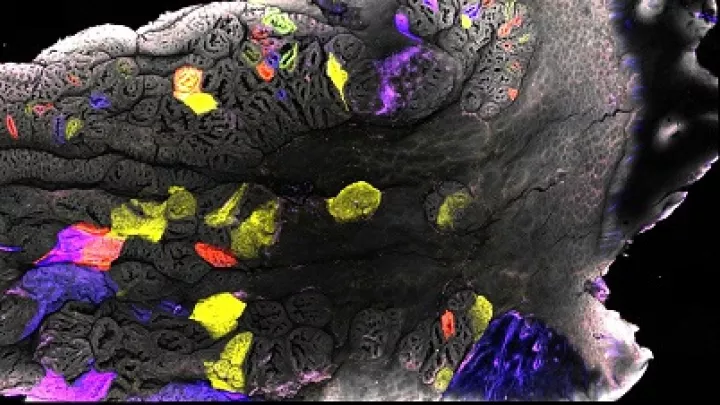
“The system we’re developing will enable rapid and accurate assessment of lung epithelial health by medical branch personnel in the field,” explained principal investigator David Warburton, MD, who is director of the Developmental Biology and Regenerative Medicine Research Program at The Saban Research Institute at CHLA. “Our goal is to be able to rapidly determine, not only the severity of any damage to the lung epithelium but, more importantly, which specific cells in the lung epithelium have been injured.”
Existing endoscopic lung surveillance programs rely on high-resolution white-light endoscopy, and current imaging does not provide any insight into tissue metabolic status, which complicates the assessment of acute lung injuries and lung health. The research team’s proposed enhanced visualization technology will improve the success rate and capabilities of existing endoscopic instruments by providing medical doctors with insight into cellular activity within the lung.
“The technology we develop will not only be useful to monitor and preserve the lung health of young people serving in the military, but will also be useful in miniaturized form to do the same thing in babies and children,” added Warburton. “What we discover through our scientific innovation at CHLA is often useful for children as well as older people including young adults.”
The collaboration also includes Scott E. Fraser, PhD, director of the Translational Imaging Center at USC Viterbi School of Engineering and USC Dornsife College of Letters Arts and Sciences, and Rex Moats, PhD, director of the Translational Biomedical Imaging Laboratory at The Saban Research Institute at CHLA and an assistant professor of Radiology and Pathology, Biomedical Engineering, USC.
This work was supported by the Department of Defense, Peer Reviewed Medical Research Program through the topic of Acute Lung Injury/Respiratory Health under Award No. W81XWH-16-1-0253.