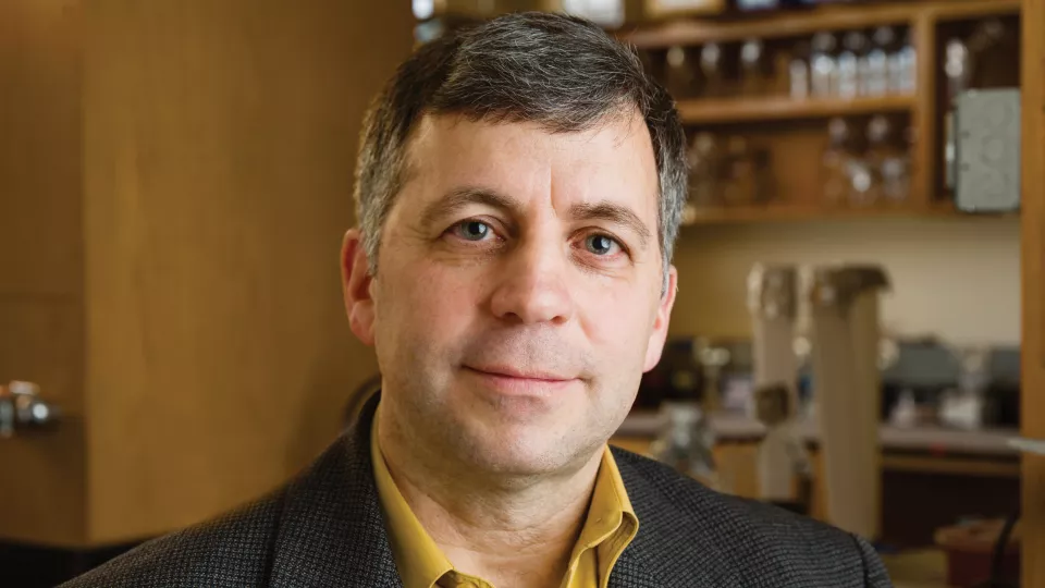
David Cobrinik, MD, PhD
Children’s Hospital Los Angeles Researcher Recognized for Contributions in Retinoblastoma Research
Retinoblastoma is a rare pediatric cancer, with approximately 250-300 new cases per year in the United States and 8,000 worldwide. The cancer grows within the retina, a thin layer of cells at the back of the eye, and is usually treatable when diagnosed early. However, if undiagnosed, retinoblastoma can metastasize and lead to death. Research has led to remarkable advances in the understanding of this disease over the past decade. David Cobrinik, MD, PhD, has contributed to many of these advances in his work at the Vision Center at Children’s Hospital Los Angeles. Recently, Dr. Cobrinik was invited to write an article about the current understanding of the development of retinoblastoma. The article appears today in The New England Journal of Medicine.
Retinoblastoma occurs when there is a specific genetic mutation in a gene called RB1, a tumor-suppressing gene. RB1 tells the body to make a protein called pRB. Since the gene was identified (it was first cloned for study in the 1980s by CHLA researchers and others), research has revealed much about how RB1, and pRB, suppresses tumorigenesis. “pRB basically puts the brakes on cell growth,” says Dr. Cobrinik, Principal Investigator in Ophthalmology at The Saban Research Institute. “However, further study of the pRB protein showed that it is normally expressed in almost all the cells of the body, so it was a mystery why children who carry an inactivated RB1 gene mainly develop tumors in the retina.
The Cobrinik Lab found that the retinal cell of origin for retinoblastoma is the cones—the color-sensing cells in the back of the eye. They then showed that the cancer occurs only when the cones are at a specific, intermediate stage of development—after the cones are immature but before they are fully developed and used for vision. They found that these “maturing cones” are extremely sensitive to RB1 gene mutations. This was one of the first demonstrations that cancers can begin in a specific stage of cell development.
Their studies also showed that these cone cells form something called a premalignant lesion, considered a dormant precursor to cancer. This lesion can then convert into a cancer from the time the baby is born up until about 5 years of age. Moreover, after the retinoblastomas form, they acquire additional mutations that make the cancers more aggressive, harder to treat and more likely to metastasize.
All of the new understanding about how retinoblastomas develop has uncovered potential therapeutic targets for possible future treatment of the cancer. It has also coincided with the development of a new biopsy method that allows doctors to determine the stage of retinoblastoma before treatments begin, which was developed by the CHLA ocular oncology team of Jesse L. Berry, MD, and Liya Xu, PhD. The expectation is that this new understanding of how tumors form combined with new diagnostic approaches will significantly improve retinoblastoma outcomes, especially for patients whose retinoblastoma tumors are only first detected in advanced stages.
For his numerous contributions to the understanding of the disease, Dr. Cobrinik was recently recognized by the International Society for Genetic Eye Disease and Retinoblastoma. “I felt very fortunate and honored to give the award lecture last summer,” says Dr. Cobrinik. “We’ve learned a lot about this disease. And we can now envision a new paradigm for retinoblastoma treatment in the future.”
Dr. Cobrinik’s work is funded by NIH grant R01CA137124, the Neonatal Blindness Research Fund, the A. B. Reins Foundation, and the Knights Templar Eye Foundation.