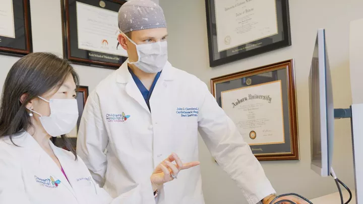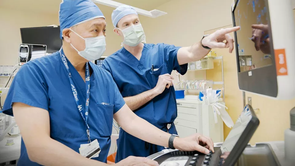
L-R: Pierre Wong, MD, and Luke Wiggins, MD
Achieving Excellence in Infant Mitral Valve Repairs
For babies and children with congenital mitral stenosis, it’s well-established that repairing the mitral valve leads to better outcomes than replacing it. But repairing the valve is not always possible—and success requires a highly integrated team approach.
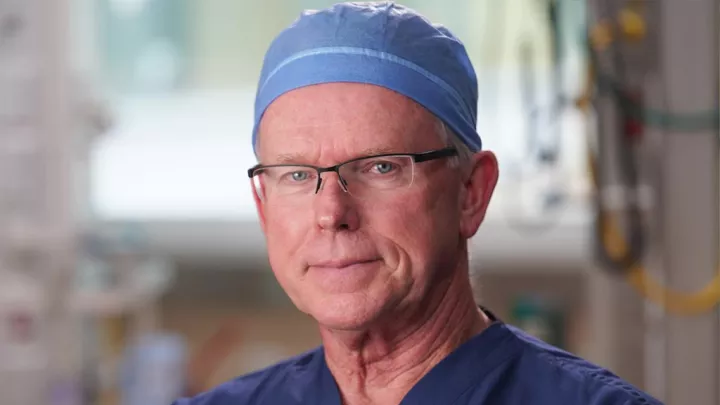
The Heart Institute at Children’s Hospital Los Angeles has extensive experience in performing the most complex mitral valve repairs in infants and children. Below, pediatric cardiologist and Director of Echocardiography Pierre Wong, MD, and pediatric cardiothoracic surgeon Luke Wiggins, MD, discuss the team’s preoperative planning process, their keys to success—and what the future may hold for mitral valve repairs.
What is the biggest challenge in congenital mitral valve stenosis?
Dr. Wong: The mitral valve is literally one of the most complicated structures in the heart. The valve itself has so many levels. The bottom line is that there’s an obstruction of blood flow from the left atrium to the left ventricle. But there are many different things that can cause that obstruction. It’s very anatomically heterogenous.
Dr. Wiggins: Every child is a little bit different. That’s why preoperative planning is so critical. It’s very important for us to understand each valve’s particular constellation of anomalies so we can better predict if we can successfully repair the valve once we get into surgery.
What preoperative imaging are you using and why?
Dr. Wong: An echocardiogram is the best way to evaluate the mitral valve because you can see the valve working in real time. The most important thing is to characterize the valve as much as possible—not just the distinct anatomical components, but also the size and function of the valve as a whole.
We use a variety of different methods with echocardiography, including 3D echocardiography, to help the surgeons understand the anatomy of the valve, the motion of the valve and what is the likelihood of success with a valve repair.
Dr. Wiggins: As Dr. Wong said, an echocardiogram is the gold standard for valve imaging. Cardiac MRI is also an important imaging adjunct to keep in mind in patients with borderline left ventricular sizes. MRI isn’t as good at looking at the mitral valve itself, but it helps us evaluate the size and function of the left ventricle to determine the suitability for biventricular circulation in very young patients.
How do you assess the severity of the stenosis? What specifically are you looking for?
Dr. Wong: We’re looking at the size of the valve, the level of the obstruction—is it one or more levels? What is the nature of the stenosis itself? If it’s a membrane that’s above the valve, that will be easy to take out and less of a problem. But if the valve is very thick and portions are fused together or poorly differentiated from one another, it might be much more difficult for the surgeons to create a competent valve out of that.
We have to look at every level of the valve—the area above it, the leaflets, the chordae, the papillary muscles. Is there a significant obstruction across the valve that is causing dilation of the left side and elevation of the left atrial and pulmonary artery pressures? Are there other associated problems with the left ventricle? Is the aortic valve diseased, or is there something else going on that might require surgery? It’s a complex and comprehensive process.
Dr. Wiggins: One of the most important aspects of the preoperative workup is to discuss the timing of the intervention, as well as the potential for repair. We really have to strategize the appropriate timing of attempting a valve repair, with the understanding that a valve replacement may be indicated once we get into surgery. Surgical timing is unique to each patient. Mitral valve disease in the pediatric population requires an individual strategy for each patient to optimize valve function and minimize the number of mitral valve procedures over a patient’s lifetime.
How has your approach to congenital mitral valve stenosis evolved?
Dr. Wiggins: The complexity of our repairs has really increased in recent years. It’s been proven now that mitral valve repair, especially in the pediatric population, has superior patient outcomes in the short and long term.
And so we’re much more aggressive at trying to repair these valves than we were five, 10 years ago—intervening on the subvalvular apparatus, removing supervalvular membranes, and patching and thinning the valve leaflets themselves.
Dr. Wong: It’s a very artful type of procedure the surgeons are doing. They really have to tailor the repair to what they see in the anatomy, and the anatomy differs a lot from patient to patient. And while we have excellent imaging, the surgeons can’t make a final decision until they inspect the actual valve. That’s where our surgeons’ vast experience is so important.
What do you think the future holds for pediatric mitral valve repair?
Dr. Wong: From an imaging standpoint, as we get higher-resolution imaging, both from a spatial and temporal standpoint, that will further improve our understanding of mitral valve pathology and motion. There are some efforts out there to do 3D modeling of mitral valves, but that’s still static. You need a dynamic model of the valve. But I think that’s coming with time.
Dr. Wiggins: Improvements in imaging and our understanding of the valvular pathology will help us further refine the way we repair the valve. There are many techniques that can be honed to improve these valve repairs, make them last longer and implement them in more patients.
An important pillar of advancing that care is research, which is something our team is in the process of expanding. We want to continue to enhance our ability to predict whether we will be able to repair the valve.
What is the key to the Heart Institute’s success in mitral valve repairs?
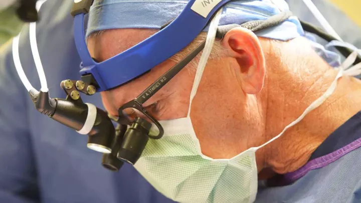
Dr. Wong: We have a long history of excellence and innovation with all types of pediatric valve surgery and valve management here at Children’s Hospital Los Angeles. In addition to mitral valve repairs, Vaughn Starnes, MD, and his team of surgeons have been leaders with the neonatal Ross procedure with aortic valve replacements, as well as with the Starnes procedure in Ebstein anomaly.
We have depth in so many areas, and that’s critical for these complex repairs. It’s vital for patients to be at a high-volume center like ours where there’s a breadth and depth of expertise across multiple disciplines.
Dr. Wiggins: The multiple disciplines are key. We have a fantastic collaborative approach. Each member of the team, from each discipline—cardiology, cardiothoracic surgery, imaging, cardiac catheterization, cardiac anesthesiology, cardiac critical care and more—come together to contribute their own piece of the puzzle. We all have the same goal: to provide the best possible care and give each child the best and healthiest future.