Research Topics
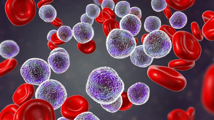
- Acute lymphoblastic leukemia (ALL)
- Childhood cancer treatment
- Immunology
- Stem cell biology
- Cancer cell biology
Research Overview
Resistance to chemotherapy and resulting relapse of cancer remains a major problem. Current treatments neglect the role of the bone marrow, which is known to allow cancer cells to survive chemotherapy. Exactly how the bone marrow can act as a safe haven sheltering cancer cells from chemotherapy is unclear.
Our lab investigates the interactions between bone marrow and leukemia and how to overcome drug resistance by disruption of such interaction. For this purpose, we established cutting-edge imaging approaches used to dissect the interaction of drug resistant leukemia cells and their protective tumor microenvironment.
Our studies are critical to understanding drug resistance and will lay the groundwork for developing effective, new therapies. Positive findings can be translated to other cancers if similar drug resistance pathways are shared.
1. Novel imaging studies of the microenvironment of drug resistant leukemia
Our understanding of drug resistance is hampered by static histological images. We have established now cutting-edge imaging approaches allowing us to study drug resistance in real time. The overall goal is to use our novel imaging approaches to dissect further the interaction of drug resistant leukemia cells and their protective tumor microenvironment.
2. Understanding integrin-mediated adhesion of ALL cells
The bone marrow niche has been found to be chemoprotective for leukemia cells, contributing to the lack of efficacy of treatment. Thus, dislodging leukemia cells from their protective niche by breaking adhesive bonds (anoikis) may make drug treatment more effective. We have identified integrin alpha4 as a central adhesion molecule in drug resistance (Hsieh et al, Blood, 2013; Shishido et al, Front of Oncology, 2014; Hsieh et al, Leukemia, 2014).
3. Apoptosis: Survivin
Survivin/BIRC5, an inhibitor of apoptosis (IAP) protein is selectively expressed in fetal and proliferating tissues and is critical for the survival and proliferation of various solid tumors and hematological malignancies. In addition, elevated survivin expression in cancer has been associated with poor prognosis. In particular, gene expression analysis of matched diagnosis-relapse pairs of ALL samples revealed higher expression levels of survivin at relapse than at diagnosis (Park et al, Blood 2011). Therefore, we aim to evaluate inhibition of survivin as a therapeutic target in ALL.
4. Self-renewal: Wnt/beta-catenin signaling
While survivin appears to play an important role in the initiation and progression of various human cancers, its mechanism is not explained fully. A better understanding of both the physiologic and pathophysiologic roles of survivin is critical for further clinical application of reagents that inhibit survivin expression. Earlier studies by Dr. Michael Kahn (USC) in colon cancer cells suggested that the regulation of survivin expression is at least partially WNT/β-catenin dependent. (Emami et al, 2004). Our hypothesis is that inhibiting the transcription of the survivin gene, via disrupting the CBP/β-catenin interaction, will lead to selective elimination of drug-resistant ALL cells (Gang et al, Oncogene 2014).
Funding
- NIH R01CA172896
- St. Baldrick’s Foundation Scholar
- Hyundai Hope on Wheels Scholar
- V-foundation
- William Lawrence and Blanche Hughes Foundation
- Couples Against Leukemia
- Whittier Foundation
- Nautica