A Higher Resolution Image of Human Lung Development
The invention of interactive map applications has revolutionized wayfinding, providing an unprecedented level of information far beyond what printed road maps can offer. Researchers at Children’s Hospital Los Angeles are giving us a similar look into the anatomy of the human lung, and their findings could help babies breathe easier.
Infants born prematurely often suffer from poor lung development and can face life-threatening consequences. In order to provide novel treatments for these babies, we must first understand how cells of the lung differentiate and grow. But there are large knowledge gaps that must first be filled. Denise Al Alam, PhD, leads a team that studies development of the lung on a molecular and cellular level.
A new study, published online in European Respiratory Journal, marks an important milestone. “This is one of the very first studies looking at how the human lung develops at the single cell level,” says Dr. Al Alam. The study tracked the fate of these cells over time, showing a trajectory of cell development. Knowing when certain cell types are differentiating gives investigators a very detailed, time-lapse map to lung development.
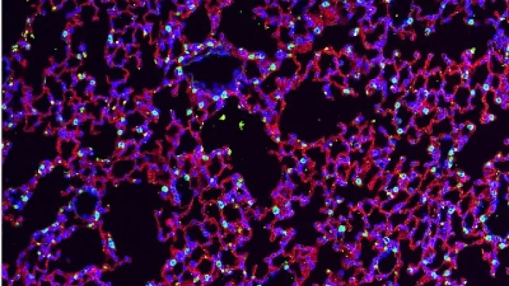
Dr. Al Alam’s team focused on the development of two types of cells in the lung. Airway smooth muscle cells line structures such as the trachea, bronchial tubes, and smaller branches. Vascular smooth muscle cells are found in the walls of blood vessels. Both types of cells add muscle tone and stability while also mediating necessary movement within these structures. But they are often implicated in different disease pathways.
“The problem is that we can never really study one cell type without also studying the other,” says Dr. Al Alam, investigator in the Developmental Biology and Regenerative Medicine Program at The Saban Research Institute of CHLA. Up until now, investigators have used a marker – known as ACTA2 – to identify these cells. But the marker doesn’t distinguish between the two smooth muscle cell types. Although both types of cells are smooth muscle they are implicated in totally different pulmonary diseases. “We need to study the two types of cells independently to understand what happens in different lung disorders.” Fortunately, her work has solved this problem that has hindered research in the field.
Dr. Al Alam’s team has identified molecular markers that are unique to each subset of cells. This will allow investigators to differentiate between the two groups, which will benefit respiratory research efforts worldwide. Developmental disorders such as COPD, asthma, and bronchopulmonary dysplasia can now be studied separately from related, but distinct, conditions that arise later in life, such as pulmonary hypertension.
“It’s been decades since we’ve introduced anything into the clinic to treat lung diseases associated with prematurity,” says Dr. Al Alam. “We are putting together critical pieces so we can get these babies the treatments they need.”
The first author of the study was Soula Danopoulos, of The Saban Research Institute of CHLA. Other authors on the study were Soumyaroop Ghattacharya and Thomas J Mariani of the University of Rochester. The study was funded by the National Institutes of Health National Heart, Lung, and Blood Institute (R01HL141856, R01 DA037447) and the Hastings Center for Pulminary Research.