
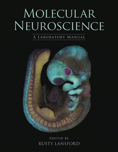
Rusty Lansford, PhD
Dr. Lansford is the recipient of the NASA Space Act Award for his contributions of Two-photon Microscope Imaging Spectrometer for Multiple Fluorecent Probes. Lansford has multiple patents for his systems and methods for monitoring cellular activity, as well as for his work in laser microscopy and imaging spectrometry. This development of a multispectral imager that allows the emission spectrum to be acquired from a single scan of specimen led to a multispectral detector based upon his invention, which is being sold by Zeiss.
Dr. Lansford’s scientific contributions, have been featured in the Pasadena Museum of California Art in an exhibit entitled, “Data + Art: Science and Art in the Age of Information,” as well and the San Francisco Exploratorium and California Science Center, where his Quail Developmental Atlas procured from basic research as a biology, math, and physics educational tool.
Accomplishments
The form and function of embryogenesis and pathogenesis
Many diseases can be considered as a failure of development. Investigating the borderland of embryology and pathology is a fundamental method for learning the rules of development. My group investigates the fundamental principles that guide how cells self-organize through collective interactions to bring about changes in embryonic form and function. We are interested in how molecules work together to control the timing and the spatial pattern of cell differentiation in developing tissues and stem cell systems. We take an interdisciplinary approach to understand the relationships between chemical, genetic and mechanical cues that shape the developing embryo.
Publications
Recent Publications
Bénazéraf B, Beaupeux M, Tchernookov M, Wallingford A, Salisbury T, Shirtz A, Shirtz A, Huss D, Pourquié O, François P, Lansford R. Multiscale quantification of tissue behavior during amniote embryo axis elongation. Development, in press, 2017. [Epub ahead of print; doi: 10.1242/dev.150557]
Sato Y, Nagatoshi K, Hamano A, Imamura Y, Huss D, Nomura T, Uchida S, Lansford R. Basal filopodia and vascular mechanical stress organize fibronectin into pillars bridging the mesoderm-endoderm gap. Development, 144: 281-291, 2017.
George L, Dunkel H, Hunnicutt J, Filla M, Little C, Lansford R, Lefcort F. In vivo time-lapse imaging reveals extensive neural crest and endothelial cell interactions during formation of the peripheral nervous system. Dev Biol. 413:70-85, 2016.
Huss D, Benazeraf B, Wallingford A, Filla M, Yang J, Fraser SE, Lansford R. Transgenic quail to dynamically image amniote embryogenesis. Development. 142:2850-2859, 2015.
Mahadevan A, Welsh IC, Sivakumar A, Gludish DW, Shilvock AR, Noden DM, Huss D, Lansford R, Kurpios, NA. The left-right Pitx2 pathway drives organ-specific arterial and lymphatic development in the intestine. Dev Cell. 31:1-17, 2014.
Aleksandrov A, Czirok A, Szabo A, Filla MB, Hossain MJ, Whelan PF, Lansford R, Rongish BJ. Convective tissue movements play a major role in avian endocardial morphogenesis. Dev Bio. 363:348-61, 2012.
Bower D, Sato Y, Lansford R. Dynamic lineage analysis of embryonic morphogenesis using transgenic quail and 4D multispectral imaging. Genesis 49:619-643, 2011.
Sato Y, Poynter G, Huss D, Filla MB, Rongish BJ, Little CD, Fraser SE, Lansford R. Dynamic analysis of embryonic vascular development in transgenic quail. PLoS One 5:1-12, 2010.
Research
The Lansford research group investigates the fundamental principles that guide how cells self-organize through collective interactions to bring about changes in embryonic form and function, with our particular focus in the developing neural and vascular systems. We are interested in how molecules work together to control the timing and the spatial pattern of cell differentiation in developing tissues and stem cell systems. Over the past decade we developed transgenic, fluorescent protein (FP) expressing Japanese quail as an experimental system. We simultaneously developed state-of-the-art live cell and tissue imaging methodologies and use them to better understand the complex cellular processes underlying embryonic development and disease. The transgenic quail model system is rapidly emerging as we share our transgenic lines prior to publication and teach labs worldwide how to work with the tools and techniques of imaging and genetically perturbing their embryos.
Visit the Lansford Laboratory.
Current Funding
The Lansford lab thanks the generous support of the Human Frontiers Program, the National Institute of health, the Rose Hills Foundation, and the CHLA Foundation.
Awards
NASA Space Act Award for Two-photon Microscope Imaging Spectrometer for Multiple Fluorescent Probes (along with Greg Bearman and Scott Fraser) 2003
R&D 100 Award for development of META multispectral imager (along with Greg Bearman, Scott Fraser, and Carl Zeiss Jena GmbH) 2002
Bridge Art+Science Alliance Award "New Ways of Seeing the Pattern: Exploring Heart Formation in a Gestural Interface System" (along with John Carpenter) 2017
Resources
Society of Developmental Biology
Eli and Edythe Broad Center for Regenerative Medicine and Stem Cell Research at USC